Technical Support技术支持
CONTACT US
 400 179 0116
24-hour service hotline marketing@ldraft.comE-mail
Literature summary: How to conduct cloning experiments
source:QiDa technoligy views:2893 time:2023-05-19
Clonogenesis assay is a useful tool for testing whether a given cancer treatment can reduce the clonogenesis survival rate of tumor cells. Colonies are defined as clusters of at least 50 cells and can usually only be identified under a microscope. Clonogenesis tests are the preferred method for determining cell reproductive death after ionizing radiation therapy, but can also be used to determine the effectiveness of other cytotoxic drugs. The following scenario has been modified from the published version (Franken et al., 2006).
Materials and reagents
Cell culture-medium
Phosphate buffered brine (PBS) (Qida Biological, article number: SD0029)
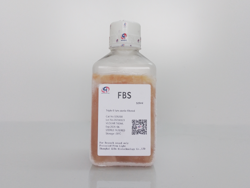
Fetal Bovine Serum (Qida Bio, item No. : SD0200)
Trypsin /EDTA
Crystal violet


equipment
Cell culture dishes or six-well plates
hemocytometer
Stereoscopic microscope
hemocytometer
thermostat
Operation procedure
1. Select the cells you need to culture according to the references.
2. Remove the culture-medium and rinse the cells with 10ml PBS.
3. Add 4ml 0.25% trypsin to the cells and incubate at 37°C for 1-5 min until the cells are rounded.
4. 10ml of medium containing 10%FBS was added and the cells were separated by pipetting.
5. Use a blood cell meter to count your cells.
Note: Getting a relatively accurate cell count is crucial.
6. Prepare the desired inoculation concentration and then inoculate the cells into a petri dish or 6-well plate.
Electroplating before treatment:
1. Harvest the cells and grow the appropriate number of cells in at least two copies on each petri dish or each well on a 6-well plate. The number of cells used for inoculation should be determined according to the aggressiveness of the treatment. The cells were grown in a CO2 incubator at 37°C for several hours and attached to a culture plate/dish.
2. Treat cells with chemicals, radiation (such as 2-10Gy), or a combination of both if necessary.
3. The cells were cultured in a CO2 incubator at 37°C for 1 to 3 weeks until the cells in the control plate formed basically good sized colonies (50 cells per colony was the minimum score).
Treated coating:
1. Harvested cells after treatment. Can plate 50 cells or up to 50 x 104 cells. If the treatment effect is unclear, prepare a series of diluents with different numbers of cells. For radiation therapy, cells can be inoculated immediately after treatment or re-inoculated later. It is best to leave the cells on ice before reinoculation.
2. The cells were cultured in a CO2 incubator at 37°C for 1 to 3 weeks until the cells in the control plate formed basically good sized colonies (50 cells per colony was the minimum score).
Fixation and staining:
1. Remove the medium and rinse the cells with 10ml PBS.
2. Remove PBS, add 2-3 ml fixing solution, and place petri dish/culture plate at room temperature (RT) for 5 min.
3. Remove the fixing solution.
4. Add 0.5% crystal violet solution and incubate at room temperature for 2 hours.
5. Add 10ml of medium containing 10%FBS and separate the cells by pipetting.
6. Carefully remove crystal violet and rinse the dish under running water.
7. Air dry the dishes/dishes on a tablecloth at room temperature for a few days.
Data analysis:
1. Count the number of colonies with a stereo microscope.
2. Electroplating efficiency (PE) and survival rate (SF) were calculated.
PE= Number of colonies formed/number of cells inoculated x 100%
SF= number of colonies formed after treatment/number of cells inoculated x PE
Colony fixation solution formula
Acetic acid/methanol 1:7 (volume/volume)
Crystal violet 0.5% solution以上翻译结果来自有道神经网络翻译(YNMT)· 通用场景
reference:Yang, X. (2012). Clonogenic Assay. Bio-protocol 2(10): e187. DOI: 10.21769/BioProtoc.187.


