Technical Support技术支持
CONTACT US
 400 179 0116
24-hour service hotline marketing@ldraft.comE-mail
The operation of cell phagocytosis experiment, the "Hunger games" of cells, was described
source:QiDa technoligy views:1897 time:2024-04-19
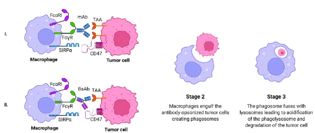
Cytophagocytosis is the function of some specific cells in the organism to recognize foreign bodies and swallow and destroy them. It's a basic biological defense mechanism
But it sounds like a cellular version of the Hunger Games.
In this game, our cells become brave fighters, and their opponents are the little bad guys - like bacteria, viruses, and other undesirable little particles.
Imagine that these cells are like city police, and bacteria and viruses are like street thugs.
When these hybrids (bacteria, viruses) try to wreak havoc in the city (our body), the police (the cells) are quick to catch them and throw them in jail (some small corner of the cell).

This process is called cytophagy. The cells stick out their "big hands" (which are actually things called pseudopods) to stick bacteria, viruses, or other small things together and then swallow them up like Pac-Man. Once these little things are swallowed, the cells begin to process them, rendering them inactive and ensuring that they do not cause further damage to our bodies.
So, cytophagocytosis is like the cellular version of cops catching thieves, except the thieves become tiny bacteria and viruses, and the cops become brave cells.
Well, let's see how this brave cytophagy experiment works.

Cytophagocytosis is a process of observing cell phagocytosis and is often used to study the immune function and defense mechanisms of cells. The following is a basic procedure for phagocytosis experiments:

Procedure of cytophagocytosis experiment
I. Preparation of experiment
1. Cell culture: Select the appropriate cell lines to ensure that they grow well in the desired medium and are in a logarithmic growth phase.
2. Reagent preparation: preparation of phagocytic substances (such as fluorescently labeled bacteria, particles or cell fragments), as well as necessary media, pancreatic enzymes, PBS, etc.
3. Equipment preparation: microscope, Transwell plate, cell counter, pipette, incubator, centrifuge. Flow cytometry, etc.
Ii. (1) Routine cytophagy experiments
1. Cell inoculation: The cells are inoculated into the upper chamber of the Transwell plate to ensure that the cell density is moderate in order to observe phagocytosis.
2. Add phagocytic substances: Add fluorescently labeled phagocytic substances to the upper chamber of the Transwell plate to ensure that the substances are evenly distributed around the cells.
3. Cell culture: Put the Transwell plate into the incubator and culture according to the needs of cell growth.
4. Fixation and staining: After the culture, take out the Transwell plate, fix the cells with appropriate fixation solution, and then stain the nucleus with a staining agent (such as DAPI).
5. Observation and recording: Observe cell phagocytosis with fluorescence microscope, and record the quantity and distribution of phagocytosis substances.

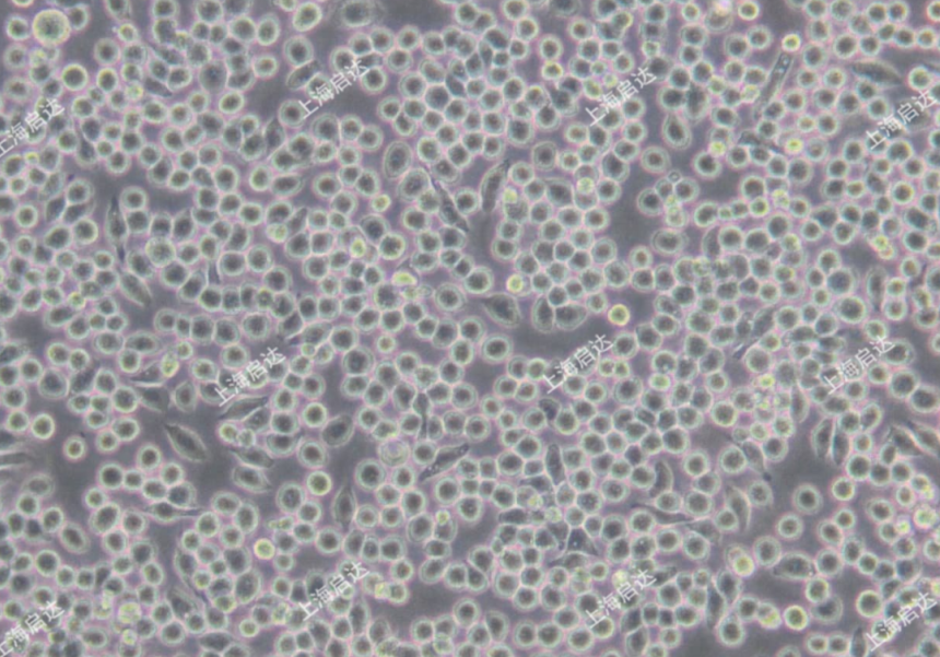
png J774A.1 (Keida Biology, item No. : CD0211)

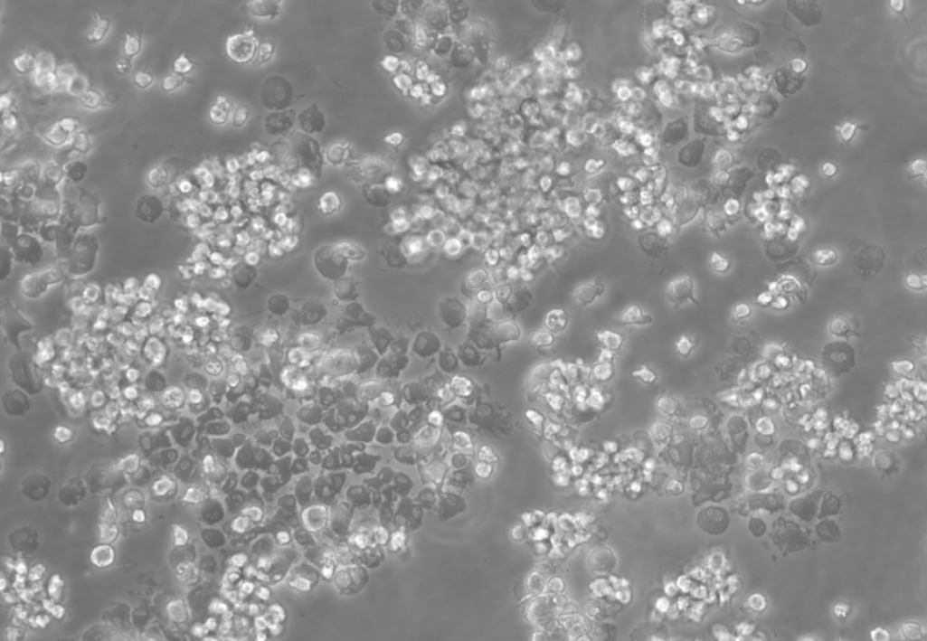
NR8383 (QIDA Biology, Code: CD0152)
(2) Operation of phagocytic cell adhesion and phagocytic rate of bacteria: treatment group, control group and cytorelaxin D treatment group were treated with 20ug/ml cell concentration of 1*10^8/mL, CFDA-SE working concentration of 5μM, incubated at room temperature and away from light for 30min, and gently mixed inversely once /10min during incubation.
(3) Bacteria interact with cells. The prepared bacteria in step (2) are incubated with phagocytes of each group. After incubation, the control group is incubated with 0.4% Trypan blue at room temperature for 5min;
(4) Flow cytometry, the control group was used as the gate setting standard in the detection, the detection result of the untreated group was the sum of the adhesion rate and phagocytosis rate, and the detection result of the cytorelaxin D treatment group was the adhesion rate. Thus, the adhesion rate and phagocytosis rate of phagocytes to bacteria were calculated.
3. Data analysis and result interpretation
1. Data analysis: counting the number of phagocytic cells, calculating the phagocytic rate, and analyzing the distribution and quantity of phagocytic substances.
2. Interpretation of results: According to the results of data analysis, explain the ability of cells to phagocytosis and the possible influencing factors.
4. Precautions
1. Aseptic operation: aseptic operation should be maintained during the experiment to avoid contamination.
2. Cell state: Ensure that cells are in a good state of growth and avoid using aging or sick cells.
3. Reagent quality: Use high-quality reagents to ensure the accuracy of experimental results.
4. Use of instruments: Use instruments correctly and avoid long-term exposure to ultraviolet light to protect cells from damage.
Such a simple phagocytosis experiment, have you learned it? You can leave a message in the background, add our wechat, and teach the technology to do the experiment


