Technical Support技术支持
CONTACT US
 400 179 0116
24-hour service hotline marketing@ldraft.comE-mail
Master the following 9 tips to easily isolate and culture epidermal keratinocytes
source:QiDa technoligy views:2152 time:2024-04-18
Master the following 9 tips to easily isolate and culture epidermal keratinocytes
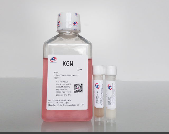
Prepare KGM media in advance (for the first extraction, 2.5% horse serum is recommended to prevent keratinocyte differentiation)
The medium is as follows, a serum-free component with no animal source protein, 500ml set, easy to use

Prepare the buffer solution for rinsing tissue in advance by pre-cooling. Our recommendation is to store it in the refrigerator at 4° after preparing 50%MEM+50%HBSS+2%PSN.
Ophthalmic scissors, ophthalmic tweezers, ice bath pre-sterilized ready, 0.25%EDTA pancreatic enzyme ready
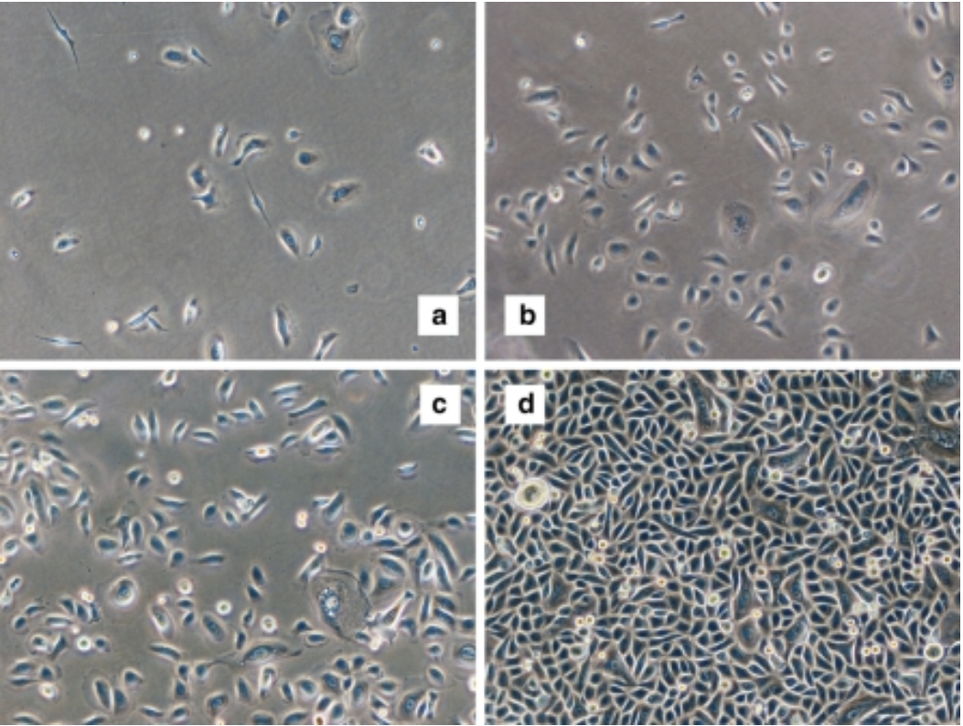
All right, so much talking, here we go. Let's take a look at our extracted cell pictures:

7-day cell growth maps of human keratinocytes extracted using KGM keratinocyte culture-medium (Keida Bio, article No. P8001)
How did you feel after reading the picture? So let's start the extraction process

The separation steps of epidermal keratinocytes mainly include the following steps:
1. Take a 1.5mm thick skin sheet and (preferably not more than 12 hours) rinse the obtained skin material with a mixture of 4℃MEM and HBSS for 2-3 times. The ice bath is done in a petri dish. The first time, the blood is flushed away, the second time, the remaining tissue is scraped away, leaving the dermis and epidermis, and the tissue is transferred to a new dish after the first rinse
2. Smooth the skin, cut it into 2-3mm² pieces with a scalpel, and put it into a neutral protease digestion solution for 15-20h at 4℃, or 2-4h at 37℃.
3. Separate dermis and epidermis with sterile needles and ophthalmic tweezers. The epidermis is thin and transparent, and the dermis is thick and slightly swollen and gelatinous under digestion by pancreatic enzymes.
4. The isolated epidermal blocks were digested with 0.25% trypsin and oscillated at 37℃ for 10-30min until the epidermis was organized into flocculent form, and then culture medium containing 10% fetal bovine serum was added to terminate digestion.
After completing the above steps, you have already seen the dawn of successful isolation of epidermal keratinocytes.
However, it must be noted that the whole process requires delicate manipulation and a strict sterile environment to ensure the integrity and viability of the cells.
5. Gently blow the epidermal tissue with a straw to make the cells fall off the tissue mass, and then use a 100um cell filter to filter and transfer the cell suspension to the centrifuge tube.
6. Add appropriate amount of KGM keratinocyte medium into the centrifuge tube, and then centrifuge at 1000rpm for 5-7 minutes to remove digestive fluid and impurities.
7. Discard the supernatant, add new KGM medium, gently blow the cells to make them uniformly suspended. After 48 hours, observe the adhesion of cells to the wall (2.5%HS is recommended for the first time).
9. Inoculate the cell suspension into an appropriate culture vessel (such as petri dish, culture bottle or culture plate), add sufficient medium, and place in the cell incubator for culture.
In the process of cell culture, the medium needs to be changed regularly to maintain the growth environment of cells. At the same time, it is also necessary to observe the growth of cells and adjust the cell density and passage as needed.
After the above steps are completed, you can obtain isolated and purified epidermal keratinocytes for subsequent experimental studies. Please note that strict aseptic practice is required during the experiment to avoid cell contamination and experimental failure. At the same time, appropriate medium and conditions should be selected according to the experimental needs to ensure the normal growth and differentiation of cells. For example, our KGM keratinocyte medium is used for keratinocyte cells, and our ECGM endothelial cell medium is used for endothelial cells.....
In the process of cell culture, we need to observe the growth state of cells regularly (it is generally recommended to observe once every two days or once a day). Healthy keratinocytes should show the characteristics of adherent growth, regular morphology, and clear cell boundaries. If the cells are found to have slow growth, abnormal morphology, pollution, etc., timely measures need to be taken, such as replacing new media, adjusting culture conditions, passing or re-separating the cells.
Well, did you get it?


