Technical Support技术支持
CONTACT US
 400 179 0116
24-hour service hotline marketing@ldraft.comE-mail
Protocol: How THP-1 Cells Differentiate M1 and M2
source:QiDa technoligy views:5152 time:2023-06-21
THP-1 cells, a human leukemia Monocyte line that can differentiate into macrophages. This allows the study of the effects of macrophage secretions on co cultured cancer cells without direct cell contact interaction (for example, the effects on growth, drug response or gene expression). THP-1 Monocyte differentiate into polarized macrophages in vitro to study the effects of M1 and M2 macrophage populations on other cells. M1 macrophages are generally considered to inhibit tumors, M2 type macrophages are believed to have tissue repair and tumor growth promoting activities, so the bipolar differentiation possessed by THP-1 is the key to many experiments. The following are the steps for THP-1 differentiation experiments, please keep it in mind. Cell experiments are not lost.


Preparation materials:
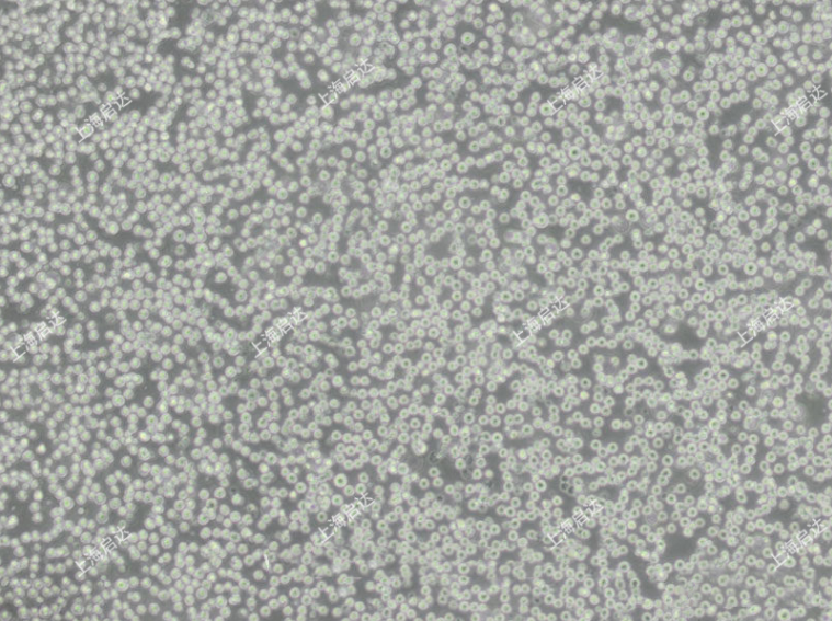
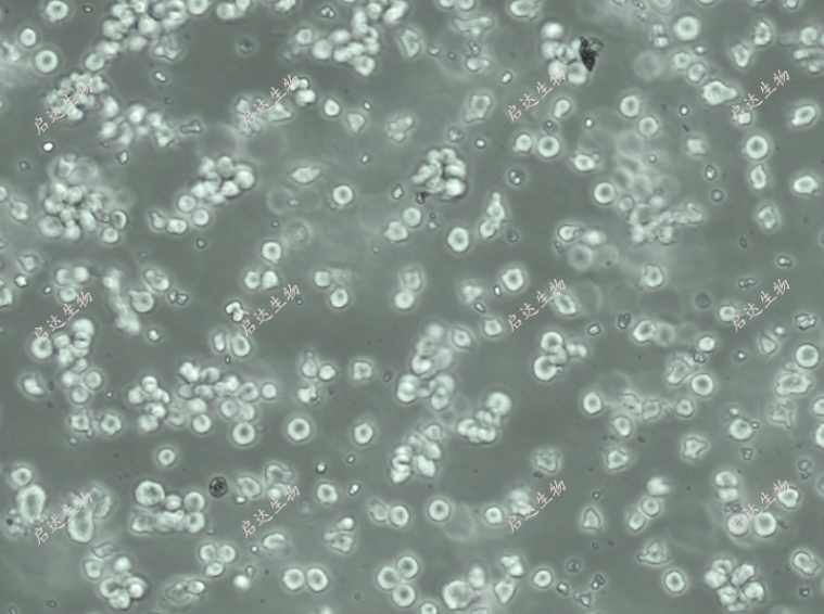
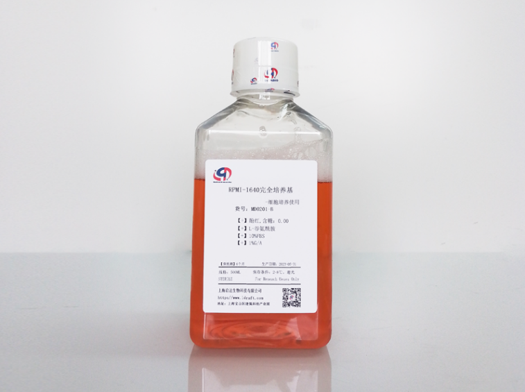
Transwell inserts 0.4 μ M. THP cells (Qida Biological product number: CD0122), 1640 normal culture medium containing 10% FBS (Qida Biological product number: MD0201), IL4, IL13, IFN- γ Reserve solution, PMA (see experimental steps), LPS (see experimental steps)

Preparation of experimental reagents:
IL4 stock solution, IL13 stock solution, IFN- γ Preparation of reserve solution: 100ug/ml was prepared in PBS containing 0.05% BSA, packaged and stored at -80 ° C; When in use: Dilute to a concentration of 40 µ g/µ l in sterile PBS, then divide equally and store at -20 ° C
Lipopolysaccharide: diluted to 40 µ g/µ l in sterile PBS, then divided into equal parts and stored at -20 ° C
Experimental steps:
Day 1:
1. THP-1 cells grow in suspension in RPMI/FCS medium and are activated once inoculated into the transwell insert. Use gloves to handle the sterile edge of the insert.
2. Use a hematometer to count THP-1 cells and obtain a concentration of 1 × 10⁵ cells/ml cell suspension.
3. Then prepare a 6-well plate by adding 2ml of RPMI/FCS culture medium every 6 wells. Then place the transwell insert into 6 wells containing 2ml of culture medium.
4. Add 2ml of THP-1 cell suspension to the insert (2x10⁵ cells/insert).
5. Once THP-1 cells were precipitated, they were immediately treated with 10 ng/ml 12-O-neneneba tetradecanoyl horbol-l3-acetate (PMA) for 24 hours (1 µ l stock solution, total volume 4 ml).
Day2:
1. THP-1 cells activated in PMA differentiated at transwell inserts 24 hours later.
2. Prepare a new 6-well plate by adding 2ml of fresh RPMI/FCS medium.
3. Then gently aspirate the culture medium containing PMA from the insert containing activated THP-1 cells and transfer the insert to a new 6-well plate
4. Then add 2ml RPMI/FCS medium to the insert and let it stand for 1 hour before treatment.
5. Then treat the insert with the required cytokine mixture:
5.1. M1 differentiation can be achieved by using 15 ng/ml Lipopolysaccharide (LPS) (1.5 µ l stock solution converted to 4 ml) or 50 ng/ml IFN- γ (2 µ l of stock solution converted to 4ml) for processing.
5.2. M2 differentiation can be achieved by combining treatment with 25 ng/ml IL4 and 25 ng/ml IL13 (1 µ l stock solution with 4 ml).
Undifferentiated but activated THP-1 cells were untreated in the insert to serve as reference wells for activated but undifferentiated Monocyte.
At this stage, correct differentiation into M1 and M2 macrophages can be evaluated by immunofluorescence, ELISA or gene expression analysis of specific markers (such as IL1, IL10, Arginase).


