

Technical Support技术支持
CONTACT US
 400 179 0116
24-hour service hotline marketing@ldraft.comE-mail
No problem! Key content: Essential steps for apoptosis operation
source:QiDa technoligy views:239 time:2026-09-01
The detection of apoptosis can be conducted from six aspects: morphology, cell membrane, DNA, mitochondria, Caspase cascade, and protein expression. The commonly used methods are flow cytometry and TUNEL staining.
Flow cytometry (Annexin V-FITC/PI double staining)
Procedure:
Collect and process the cells (including suspended cells), and wash them twice with PBS.
Resuspend the cells in 1× Binding Buffer and adjust the density to 1×10⁶ cells/mL.
Add 5 μL Annexin V-FITC and 5–10 μL PI, and incubate at room temperature in the dark for 15 minutes.
Perform flow cytometry detection within 1 hour.
Result interpretation:
Lower right quadrant (Annexin V⁺/PI⁻): Early apoptosis
Upper right quadrant (Annexin V⁺/PI⁺): Late apoptosis/necrosis
The total apoptosis rate in the model group is usually 2–3 times higher than that in the control group (e.g., from ~10% to ~30–50%).
Figure 1.png Figure 2.png
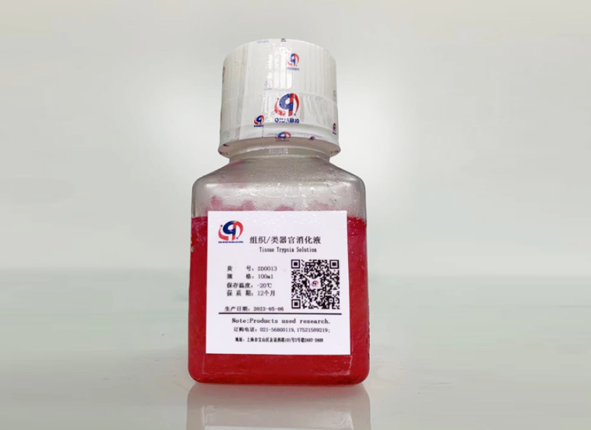
Tissue digestive enzymes (Qida Biotechnology, product number: SD0013)
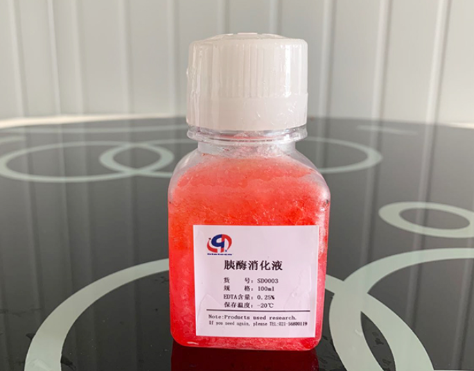
Cell digestion solution (Qida Biotechnology, product number: SD0003)
2. TUNEL staining
Procedure:
Place the cells on a 24-well plate with cell slides.
Fix with 4% paraformaldehyde at room temperature for 30 minutes.
Dip with TUNEL staining solution and incubate at room temperature in the dark for 1 hour.
封片 with DAPI, observe and take pictures with a laser confocal microscope.
Judgment: The model group shows obvious green fluorescence (TUNEL positive), while the control group has almost no signal.