

Technical Support技术支持
CONTACT US
 400 179 0116
24-hour service hotline marketing@ldraft.comE-mail
A comprehensive guide to operating endothelial cell injury models
source:QiDa technoligy views:453 time:2026-08-06
Endothelial cell injury models include: cytokine-induced injury model, hydrogen peroxide-induced injury model, oxygen free radical-induced injury model, oxidized lipoprotein-induced injury model, among others.
I. Endothelial cell stress injury model
Hydrogen peroxide (H₂O₂) is one of the most commonly used chemical inducers for establishing an endothelial cell oxidative stress injury model; it can simulate vascular endothelial dysfunction caused by excessive reactive oxygen species (ROS) in vivo. The concentration and exposure duration are critical factors determining the success or failure of this model.
Operation has begun!
Step1: Cell Culture
1.Harvest endothelial cells during the logarithmic growth phase and inoculate them at an appropriate density into a culture plate (e.g., 96-well plate: 5×10³ cells/well; 6-well plate: 1×10⁵ cells/well).
2. Culture the cells in a 37 °C, 5% CO₂ incubator until cell adhesion is achieved (typically within 12–24 hours).
Step 2: Preliminary experiments to screen for the optimal concentration
1. Set up an H₂O₂ concentration gradient: 0, 100,200,400,600,800,1000 μmol/L;
2. Set up a time gradient: 6 h, 12 h, 24 h, 48 h.
Step 3: Testing
The cell viability was assessed using the CCK-8 assay or MTT assay; the optimal concentration–time combination for achieving a viability reduction of 50%–70% was identified. At concentrations too low, cellular damage was not significant; however, at concentrations too high, irreversible necrosis occurred, which is unfavorable for subsequent drug intervention studies.
Key details to never overlook when performing the H₂O₂ experiment
H₂O₂ should be prepared fresh for immediate use: H₂O₂ is unstable and must be freshly prepared in PBS or culture medium; avoid repeated freeze-thaw cycles.
Serum effects: Catalase present in serum can degrade H₂O₂; therefore, some experiments utilize serum-free or low-serum culture media.
Cell density: excessively high densities can protect cells from oxidative damage, whereas excessively low densities may lead to complete cell death; therefore, a consistent cell density must be maintained.
Duration of action: Acute injury (30 min–6 h) primarily reflects direct toxicity; chronic injury (24 h) more closely approximates the pathological state.
Special handling for aging models: For treatment with 200 μmol/L H₂O₂ for 25 minutes, the suspended incubation method should be employed (resuspend in PBS after centrifugation, then invert and mix); following treatment, cells must be washed with PBS before inoculation and culture.
Recommended product: ECGM Endothelial Cell Culture Medium (Qida, Product No.: P1001)
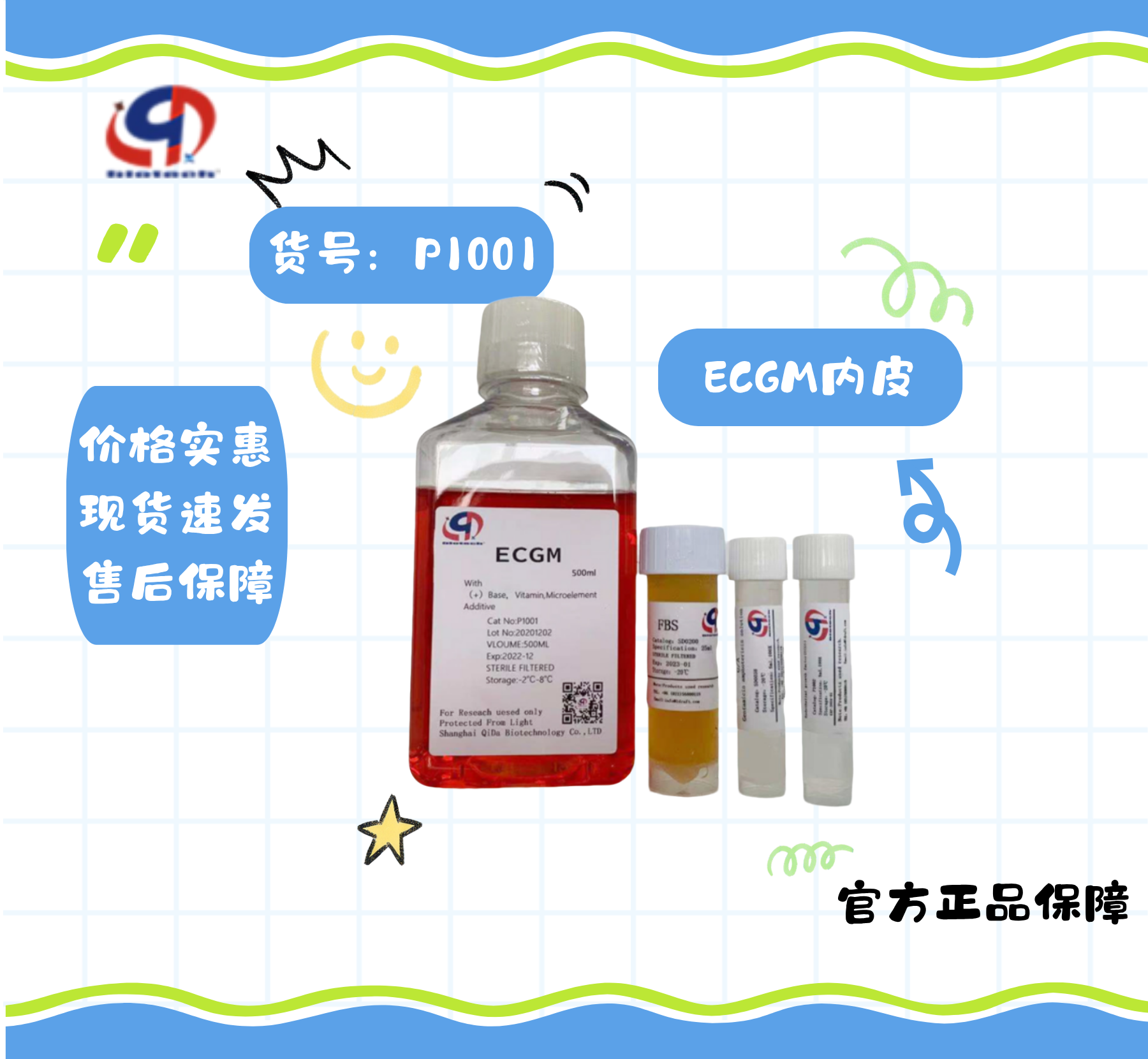
Low-serum formulation | Specifically formulated for endothelial cell growth | BBB-Preferred
Ready-to-use; ensures stable cell viability and high reproducibility of model establishment, thereby facilitating the efficient advancement of experiments.
II. Model Construction Methods for ox-LDL
1.Preparation of ox-LDL: Human plasma low-density lipoprotein (LDL; purchased from a commercial supplier or isolated in-house), CuSO₄ (analytical grade), PBS (pH 7.4, containing 0.01% EDTA), and nitrogen gas
| step | operation |
| 1. LDL Preparation | Dissolve n-LDL in PBS (without EDTA), and adjust the protein concentration to 200–500μg/mL |
| 2. Oxidative induction | Add CuSO₄ to a final concentration of 50–100 μmol/L (commonly 50 μmol/L). |
| 3. Oxidation conditions | Incubated at 37°C under shaking conditions for 16–24 h |
| 4. Termination of oxidation | Add an excess of EDTA (final concentration: 100 μmol/L) to chelate Cu²⁺. |
| 5. Dialysis desalination | Perform 4 × C dialysis (PBS, pH 7.4) for 24–48 h, with replacement of the dialysate every 6–8 h. |
| 6. Aseptic filtration | Filter through a 0.22 μm filter; aliquot and store at –80 °C. |
| 7. Nitrogen filled protection | After resealing, fill with nitrogen gas to prevent repeated freeze-thaw cycles. |
2. Steps for establishing the ox-LDL model
ox-LDL concentration selection: 80–100 μg/mL
Sample size: blank control group, model group; if drug testing is required, a positive drug control group and a drug-administration group must be prepared
Treatment duration: 24 hours
Treatment conditions: prior to treatment, switch to serum-free or low-serum (1–2% FBS) culture medium for 12–24 h, then add ox-LDL and continue incubation for 24–48 hours
ox-LDL model validation
The following assays were employed: CCK-8 assay, LDH flow cytometry, oxidative stress assay, inflammatory response assay, and endothelial function assay.
Key details to never overlook when performing the OX-LDL assay
ox-LDL source: Commercially available ox-LDL products (e.g., Sigma, Yiyuan Biotech) can be purchased; alternatively, ox-LDL can be prepared by oxidizing native LDL with Cu²⁺, though the MDA content must be measured to confirm the degree of oxidation.
Avoid serum interference: Whenever possible, use serum-free media during cell culture; antioxidants present in serum may attenuate the oxidative damage induced by ox-LDL.
Concentration sensitivity: Ox-LDL exhibits significant variability in activity across different batches; therefore, it is imperative to conduct preliminary experiments prior to the main study to screen for the optimal concentration.
Cell passage number: Excessive cell passaging may lead to alterations in cell characteristics; it is recommended to use cells from lower passage numbers.