Fibroblasts extracted using tissue adhesion method

source:QiDa technoligy views:2299 time:2023-08-17
The tumor microenvironment and cancer-related fibroblasts particularly exhibit tumor promoting ability that is not present in their normal counterparts (Calvo et al., 2013; Hanahan and Coussens, 2012). Therefore, functional and molecular characterization of the modifications that occur in fibroblasts during tumor progression is crucial for fully understanding their role in tumor progression.
1、 Method for extracting fibroblasts from breast or adipose tissue:
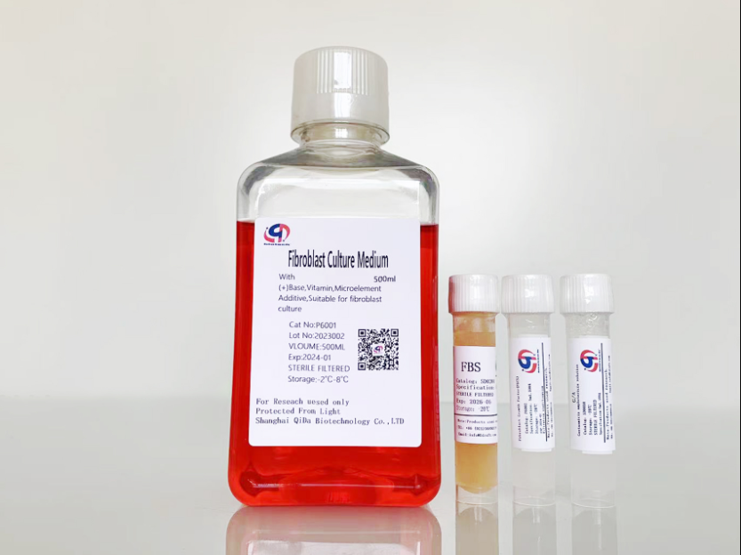
1. For normal breast tissue, cut it into 1mm fragments using ophthalmic scissors and place it in a 6CM culture dish. Then, press a 20mm cover glass slide to prevent fat tissue from floating. Then, slowly add the prepared FGM fibroblast culture medium (Qida Biological, product number: P6001), which needs to increase the serum to 10% when extracting cells. The culture medium contains small molecule compounds that inhibit the growth of heterologous cells, Adding proteins required for fibroblast growth and using FGM fibroblast culture medium can improve cell activity and purity.

The following points need to be noted when using this method:
1. The culture medium must be slowly added to avoid suspension of the slide;
2. The sample needs to come into contact with plastic and cover glass to allow fibroblasts to come out of the sample;
If a large number of fibroblasts are observed to enter the cover glass and culture dish from the tissue after
3.7 days, they can be transferred to a new culture dish.

Fibroblasts extracted using tissue adhesion method
2、Method for extracting fibroblasts from breast hyperplasia, adenoma, and cancer tissue samples:
1. Prepare a collagenase solution with a concentration of 1mg/ml and a complete culture medium containing 10ug/ml of DNase 1;
2. Place the separated tissue into a sterile tube containing an appropriate amount of collagenase/dispersing enzyme. Use a pipette to repeatedly blow several times to aid in decomposition, and then incubate the sample in an orbital oscillator at 37 ° C and 180 rpm for 60 minutes. Typically, 2-3 milliliters (250-500 milligrams of tissue) are used for each sample. Adjust according to sample size.
3. Add a volume of PBS containing 1mM EDTA to each sample and blow repeatedly several times;
4. Using a 3mm filter to filter the sample can also be done using other methods (rubber syringe plunger, etc.) or centrifuging at room temperature at 100 x g for 5 minutes (this step mainly removes undigested tissue blocks)
5. Discard the supernatant, wash the filtered cells' particles with complete culture medium, and then perform RT again with 100% × Centrifuge for 5 minutes.
6. Incubate cells containing particles with complete culture medium and DNase 1 (final concentration 10 µ g/ml) under RT for 10 minutes.
7. Repeat step 5 two to three times.
8. Discard the culture medium after centrifugation, resuspend the cells in a complete culture medium and inoculate them in a T25 or T75 bottle (usually arranged according to the number of centrifuged cells, one T25 cell should not be less than 5 * 10 ^ 5 cells), and place them in a culture incubator to begin cultivation.After
9.30 minutes, perform the first fluid change on the extracted cells using the time difference adhesion method (at this time, it needs to be replaced with a normal FGM medium containing 2% FBS). When changing the fluid, gently wash it with preheated PBS once and return it to the incubator
10. After 3-5 days of cultivation in FGM containing 2% FBS, most fibroblasts can grow to over 80%. At this time, they can be frozen or passaged according to normal methods.

Fibroblasts extracted by enzymatic digestion
Qida FGM Fibroblast Culture Medium (Qida Biology, Product No. P6001) Reference:
1.Structural and biofunctional evaluation of decellularized jellyfish matrices,DOI;https://doi.org/10.1039/D3TB00428G
2.Collagen from Iris squid grafted with polyethylene glycol and collagen peptides promote the proliferation of fibroblast through PI3K/AKT and Ras/RAF/MAPK signaling pathways,https://doi.org/10.1016/j.ijbiomac.2023.125772